Cartilage in the knee is especially vulnerable to tears from injury, particularly during sports. If you´re experiencing symptoms of a cartilage tear, Dr. Walker can help. Treatments are available for old athletic injuries, which commonly involve torn cartilage. If you have been diagnosed with a cartilage abnormality, our cartilage restoration procedures may be able to help you as well.
Dr. Walker is subspecialty board certified in Orthopaedic Sports Medicine. He can diagnose a cartilage tear by reviewing your medical history, performing a thorough examination of the joint, and ordering an X-ray and/or MRI. An MRI scan provides a very detailed picture of your joint, particularly showing the cartilage and ligaments. Dr. Walker will review with you about all of your symptoms, activities, and the circumstances that lead to the injury.
Cartilage in the joint does not have a good blood supply and has limited ability to able to heal itself, and your symptoms will get worse over time as the cartilage continues to deteriorate. Consequently, many cartilage tears require surgery for treatment.
Arthroscopic surgery is commonly used for cartilage restoration. It allows surgeons to see, diagnose, and treat problems inside a joint. Arthroscopy is less invasive than open surgical procedures. It is associated with a decreased risk of infection, minimal bleeding, less pain, and a shorter recovery period.
When possible, Dr. Walker performs cartilage repair surgery to preserve the natural knee for as long as possible and at least slow the progression of osteoarthritis, delaying the need for total knee replacement.
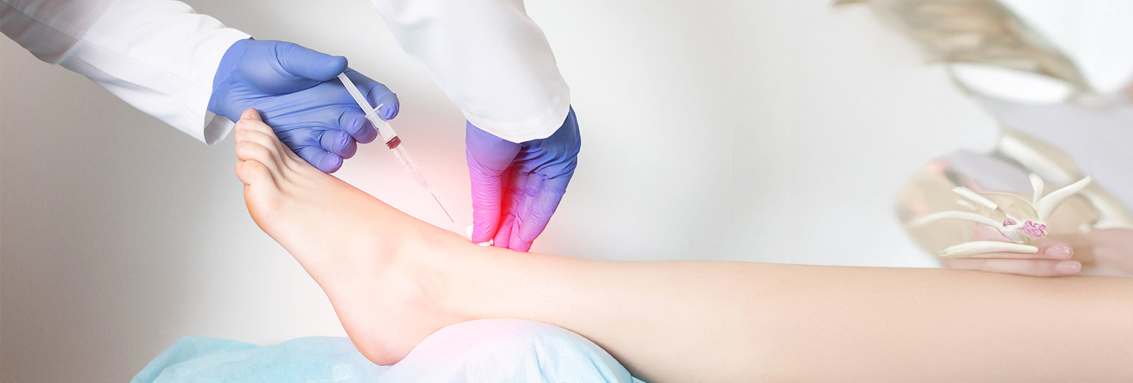
Marrow stimulation is designed to stimulate a healing response for the cartilage through a technique that accessing the bone marrow beneath the cartilage injury or defect. The marrow stimulation technique can be safe and effective in treating degenerative areas as well in early knee arthritis or cartilage injuries in active patients. This technique is performed arthroscopically for cartilage repair by stimulating stem cells from the bone marrow underneath the area where cartilage is intended to regenerate. This surgery is done as an outpatient where no overnight stay is required. Patients are asked not to put their full weight down on the joint for 4 – 6 weeks in order to keep pressure off the healing cartilage area. Range of motion and other strengthening exercises begin immediately after surgery. Studies show that the marrow stimulation techniques help improve patients´ function and reduces symptoms. With a structured physical therapy program, patients may resume low impact sports activities such as biking, swimming and walking as early as 2 months.
There are two main types of cartilage transplantation. Autologous cartilage implantation (ACI) and ostechondral articular transfer (OAT). Both techniques are FDA approved for treatment of cartilage defects in focal areas of the knee joint.
ACI is performed in two stages. The first stage is usually performed arthroscopically to harvest healthy cartilage from a nonarthritic or undamaged portion of your joint that does not support weight bearing. This is similar to removing a wall in your home that does not support the weight of the rest of the house or roof. The healthy cartilage is then processed and grown for 4 – 6 weeks to produce more cartilage producing cells. After this an open or arthroscopic surgical procedure is performed during the next stage of the technique in order to “patch” the cartilage defect in the knee. Patients are asked not to put their full weight down on the joint for 6 weeks in order to keep pressure off the healing cartilage area. Range of motion and other strengthening exercises begin immediately after surgery.
Studies show that this cartilage repair technique helps improve patients´ function and reduces symptoms. There is a well-define rehabilitation program that is utilized. Patients may resume low impact sports activities such as biking, swimming and walking as early as 3 months. Higher impact activities such as running are achievable after 12 months. Reductions in pain and improvement in function can be expected in the majority of patients treated with ACI.
Osteochondral articular transplantation or transfer is a procedure performed either arthroscopically or through a small incision on the front of the knee. It is done by transferring healthy cartilage from a lesser-weight bearing area of the knee to the portion of the knee with a cartilage defect. A small amount of bone is removed with the overlying healthy cartilage in order to “plug” in the cartilage to the site of cartilage loss. In certain situations, patients are instructed to participate in “protected” weight bearing or asked not to put their full weight down on the joint for 2 – 4 weeks in order to keep pressure off the healing cartilage area. Range of motion and other strengthening exercises begin immediately after surgery. After the procedure, a formal physical therapy program, which usually lasts 6 – 8 weeks. Patients may resume low impact sports activities such as biking, swimming and walking as early as 2 months. Running, pivoting, and other impact sports are permissible at 3 to 4 months. Studies show that the OAT technique to be an effective treatment of cartilage abnormalities. Approximately 85% of patients have good to excellent results and have improved function and reduced symptoms.
To schedule an appointment, call our Dallas office at 817.930.2030 or use our Online Appointment Request Form today. Our patients come to us from Richardson, Garland, Dallas, Fort Worth and Plano.
You won’t find a better Knee Surgeon than Dr. Torrance Walker! I’ve had too many surgeries and Dr.
Walker did my replacement a few years back. He is great and I’ve had no problems with my replacement
surgery and no problem after! I am pain free and so glad that I found Dr. Walker. He also replaced a
knee for my Mother as well as numerous friends over the years! I highly recommend Dr. Walker!
Mike Norwood
Dr. Walker did a total knee replacement on me in the summer of 2024. I am a teacher, so I get
medical procedures done during summers. Dr. Walker is the best!! You cannot go wrong having him as
your surgeon! I had the surgery on July 1st and was ready to go back to work on August 1st. By the
way, I weigh 330lbs. and it was not a problem getting back in the swing of things again.
Terence Gariano

